
Precision in dentistry is often discussed in terms of hand skills, magnification, and material science. However, the foundational element that enables all these factors to function correctly is often overlooked: light. The physics of how light interacts with the oral cavity defines the limit of what a clinician can diagnose and treat.
When we speak of visibility, we are discussing the absence of obstruction. In the deep, narrow corridors of the mouth, shadows are the silent enemy. They are not merely dark spots; they represent a total loss of visual data. If a shadow falls across a preparation margin, that anatomical information effectively ceases to exist. Understanding shadow geometry and dental illumination physics is a clinical necessity for reducing error rates.
The Physics of Shadows in Dentistry
To mitigate the impact of shadows, one must understand their anatomy. In optical physics, shadows created by a light source interacting with an object consist of distinct regions that affect visibility differently.
Penumbra vs Umbra
Every shadow has two main parts. The umbra is the fully shaded inner region where the light source is completely blocked. In this zone, visual information is zero. The penumbra is the partially shaded outer region where only part of the light source is obscured. In dentistry, the transition between these two determines edge detection. A large penumbra creates a “soft” shadow that blurs boundaries, making it difficult to distinguish where a restoration ends. Sharp, coaxial lighting aims to minimize both, ensuring total illumination of the working field.
Beam Divergence
Light travels in straight lines, but the quality of the beam depends on its collimation. Standard overhead lights often suffer from high beam diverge nce, meaning the light spreads out as it travels. This spread reduces the intensity of photons hitting the bottom of a preparation, particularly in Class II boxes. A divergent beam causes the walls of the cavity to cast shadows upon the floor. High-quality LED surgical headlights utilise lens systems to keep photons packed tightly together, ensuring deep penetration.
Angle of Incidence
The angle at which light strikes a surface determines how much illumination is returned to the eye. If light hits a molar at a steep, oblique angle, the majority of that light is reflected away from the practitioner. This results in a dark, low-contrast image. The goal of superior lighting is to align the angle of incidence as closely as possible to the visual axis, ensuring light bouncing off the tooth travels directly back to the retina.
Reflection and Refraction on Tooth Surfaces
Teeth are translucent, crystalline structures. Light enters the enamel, refracts through the dentinal tubules, and scatters. Shadow geometry becomes complex here because shadows can be cast inside the tooth structure if the light source is poor. Furthermore, specular reflection (glare) occurs when a light source is too diffuse, creating “hot spots” that wash out texture. Managing this scatter is crucial for identifying distinct anatomical features.
How Poor Lighting Alters Clinical Outcomes
When the physics of lighting is ignored, the clinical consequences are immediate. The human eye requires contrast to detect edges. Shadows reduce contrast sensitivity, forcing the brain to “guess” where a line exists.
- Misjudged Margins: The most common failure in fixed prosthodontics is an open margin. Shadows cast by the gingiva can obscure the finish line. If the bur is guided by a shadowed view, the margin may be left uneven or unsupported.
- Incorrect Shade Matching: Shadows alter the perception of value. A tooth viewed in partial shadow appears lower in value (greyer) than it is. This leads to selecting composites that are too light. Accurate color matching requires full-spectrum illumination.
- Underprepared Canals: Endodontics is a game of illumination. Inadequate light penetration prevents the location of calcified canals. If light cannot reach the pulp chamber floor due to wall shadows, the canal remains untreated.
- Missed Micro-Cracks: Early diagnosis of cracked tooth syndrome relies on seeing the refraction line within the enamel. Diffuse lighting hides these hairline fractures. Seeing clearly is the prerequisite for diagnosis, a concept explored in depth regarding the benefits of dental headlights in modern dentistry.
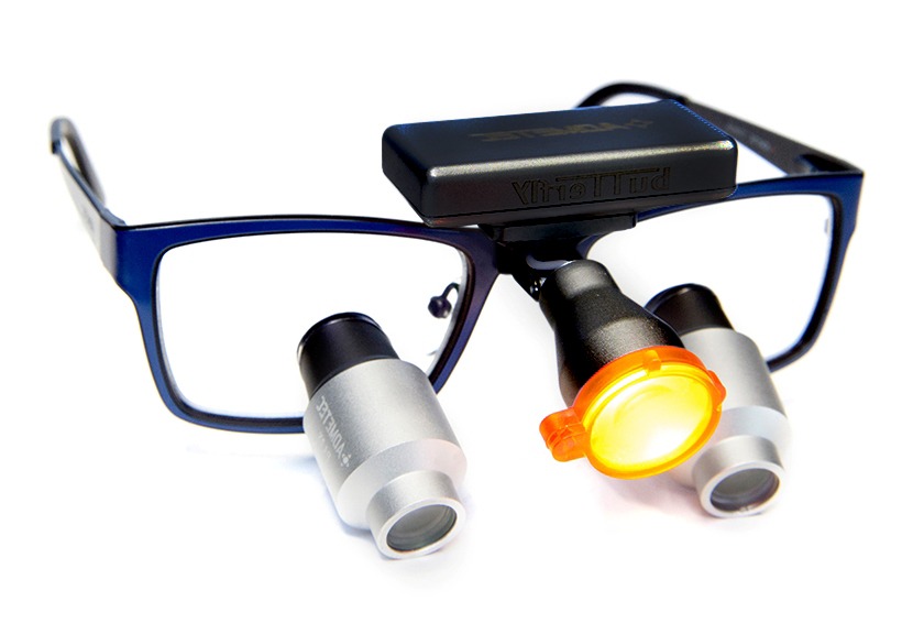
Why LED Headlights Reduce Shadow Errors
The solution to these optical challenges lies in modern technology. The shift to the portable LED surgical headlights has revolutionized visual management. This is about the geometric relationship between the light source and the target.
- Parallel Beam Geometry: High-quality headlights use collimating lenses to keep light rays parallel. This allows the beam to reach the bottom of deep cavities without being blocked by cavity walls, effectively eliminating the “bottom-of-the-well” shadow effect.
- Better Uniformity: An advanced LED system provides edge-to-edge uniformity. This means the center of the spot is just as bright as the periphery. It eliminates the “hot center” effect that causes pupil constriction and eye fatigue.
- Higher Color Rendering: The Color Rendering Index (CRI) determines how accurately we perceive the illuminated area. High CRI LEDs reveal the true contrast between necrotic and healthy dentin. For a deeper understanding of these metrics, review the factors that determine the quality of light.
How Head Positioning Changes Shadow Behavior
Even with the best equipment, physics dictates that an object placed between a light source and a target will cast a shadow. In dentistry, that object is often the dentist’s own head or hands.
- Forward Tilt: As a clinician leans forward, their head physically blocks stationary overhead lights, creating an umbra over the mouth. Headlights eliminate this by mounting the source on the head itself.
- Rotational Angle: When working on the lingual aspect of mandibular anteriors, the mirror creates shadow patterns. A light source fixed to the practitioner’s forehead rotates in sync with their view; wherever the eyes look, the light follows.
- Lateral Shifts: Shifting from 12 o’clock to 9 o’clock changes the angle of view. Static lights require manual adjustment. Head-mounted illumination corrects for lateral shifts automatically.
The Ideal Lighting Geometry for Maximum Accuracy
Achieving shadow-free dentistry requires coaxial illumination: aligning the illumination axis with the visual axis. The closer these two lines are to parallel, the fewer shadows are visible.
- Correct Beam Alignment: The light should originate from between the eyes. This ensures that any shadow cast by the hands is thrown “behind” the object of focus, rendering it invisible to the dentist.
- Correct Distance: The inverse square law states that light intensity drops off rapidly with distance. Headlights maintain a constant, optimal working distance, unlike overhead lights, which vary in intensity as the patient moves.
- Optimal Angle: The light must enter the oral cavity at an angle that illuminates the posterior regions without requiring the patient to hyperextend. This is a key differentiator when comparing dental headlights vs overhead lights.
Future Shadow-Control Innovations
The industry continues to refine the physics of medical illumination. We are approaching a point where technology may actively manage shadow geometry.
- Intelligent Shadow Prediction: Future systems may use sensors to detect obstructions and adjust intensity automatically to compensate for temporary shadows.
- Real-time Beam Correction Tech: Lenses that adapt focal length based on working distance could ensure perfect collimation at any range.
- Dual-beam LEDs: Using multiple micro-emitters at slightly different angles to “fill in” the penumbra created by instruments.
Conclusion
The pursuit of clinical excellence is a pursuit of information. Shadows act as a filter, removing critical data from the dentist’s field of view. By understanding the physics of umbra, beam divergence, and incidence angles, clinicians can make informed decisions.
Relying on static, overhead lighting introduces unnecessary variables. The geometry of a high-quality, coaxial surgical headlights system removes these variables. It provides a stable, uniform, and shadow-free environment where the only limit to accuracy is the operator’s skill. At Schultz Loupes, we engineer our lighting solutions based on these optical principles, ensuring physics works in your favor.